
What a Knee Scan Actually Shows
What an X-ray, MRI, CT and ultrasound each show in a knee, why a scan can look bad while the knee feels fine, when an MRI changes the plan and when it does not, and how a CT is used to plan robotic knee replacement.
Most people arrive at an appointment with at least one scan, and a reasonable expectation that it holds the answer. Scans are genuinely useful — but each one answers a narrow question, and none of them answers the question patients most want settled, which is what to do next.
This page explains what each type of imaging shows, what it misses, and why a report can read alarmingly while the knee it describes is doing fine.

A note on where this comes from. Before medicine, Dr Clark trained in medical imaging, holding a Master of Medical Radiation Science and a Bachelor of Applied Science in Medical Imaging. That background is directly relevant to CT-planned robotic knee replacement, where the quality of the operation is bounded by the quality of a plan built entirely from a CT dataset.
What each scan is for
X-ray
The first test in almost every knee, and for arthritis usually the only one needed.
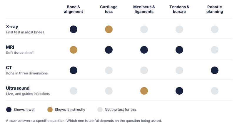
An X-ray shows bone. It does not show cartilage directly — cartilage is invisible on a plain film — so cartilage loss is inferred from the gap between the bones. As cartilage thins, the gap narrows. That narrowing, along with bone spurs, hardening of the bone beneath the joint surface, and the overall alignment of the leg, is what an X-ray contributes.
One detail matters more than most people realise: the film should be taken standing up. Joint space narrowing only becomes properly apparent when the joint is loaded. A knee X-rayed lying down can look substantially better than the same knee X-rayed standing, because without body weight through it the gap opens up. If your films were taken lying down, that is worth mentioning.
MRI
An MRI shows soft tissue — the cartilage itself, the menisci, the cruciate and collateral ligaments, the tendons, and changes within the bone that an X-ray cannot register.
It is the right test for a suspected meniscus tear, a ligament injury, or a picture that does not add up. It is often not necessary for straightforward osteoarthritis, where a standing X-ray read alongside the examination already answers the questions that determine treatment.
MRI’s sensitivity is also its difficulty. It finds a great deal, including a great deal that is not causing trouble.
CT
CT shows bone in three dimensions, with detail no other test matches.
In this practice its main use is planning. For a knee replacement planned on the Mako system, a CT scan taken beforehand is used to build a three-dimensional model of your knee, so implant size and position are decided as a virtual pre-operation before any incision is made. The VELYS system is imageless and maps the knee in theatre instead, so no planning CT is required. Which system is used depends on the hospital and theatre list.
CT is also used for complex fractures and for assessing bone stock in revision surgery.
Ultrasound
Ultrasound images tendons and bursae in real time, and unlike the others it can watch a structure move. It is the appropriate test for a suspected tendon problem or a bursitis, and it can guide an injection precisely into the right space.
It cannot see inside the joint properly, so it has little role in assessing arthritis.
The thing worth understanding about scan findings
Here is the fact that reframes most imaging reports:
Arthritis shows on the X-rays of more than half of people over 65. Only about one in seven of those have symptoms worth treating.
The same holds on MRI, more strongly. Meniscal tears, cartilage defects and bone marrow changes are common findings in people with no knee pain at all. Scan an asymptomatic fifty-five-year-old and you will usually find something to report.
This has two consequences worth taking seriously.
A scan finding is not automatically the cause of your pain. A report listing a degenerate meniscal tear, a cartilage defect and a Baker’s cyst is describing a knee that has aged. Whether any of those is what hurts is a separate question, answered by the examination and the history rather than by the scan.
A frightening-sounding report is not a prognosis. Words like “severe”, “full-thickness” and “extensive” describe appearance, not disability. Plenty of people with severe changes on paper walk comfortably, and some with modest changes are significantly limited. What the knee stops you doing is the measure that matters.
This is also why a scan cannot tell you whether to have a knee replacement. It establishes which compartments are worn — which determines whether a partial or total replacement is even an option. It does not establish whether an operation is the right choice for you. Do I Need a Knee Replacement? works through that separately.
When more imaging genuinely changes something
A scan is worth doing when the answer would change the plan. In practice that means:
- Deciding between a partial and a total replacement — which needs the state of all three compartments and the cruciate ligaments established, so CT or MRI is often added to the X-ray
- A suspected meniscus tear or ligament injury, particularly a knee that locks, catches or gives way
- A knee that does not fit the pattern — pain out of proportion to the films, rapid deterioration, night pain that is constant and unrelated to position
- Planning robotic surgery on the Mako system, where the CT is the plan
- Suspected infection or a fracture, where imaging is part of urgent assessment
And when it does not:
- Repeating an MRI that was done recently and has not changed
- Imaging to “check” arthritis already established on a standing X-ray, when the treatment question is about symptoms rather than anatomy
- Scanning a knee before trying the treatment that would be recommended regardless
What to bring to your appointment
Bring the images, not only the reports — on a disc, or the access details for the radiology practice’s online portal. A report is one radiologist’s summary; the images allow the films to be reviewed directly, which occasionally leads somewhere different.
Also bring any previous knee imaging you have, even if it is years old. Comparing a current film with an older one shows the rate of change, which is often more informative than either film alone.
If you are not yet sure what is wrong with your knee, Is It Knee Arthritis or Something Else? works through the alternatives, and Where Your Knee Hurts covers what the location of pain suggests.
This page is general information about knee imaging. It is not an interpretation of your scan, and a report should be discussed with the doctor who arranged it. If your symptoms change significantly, or the knee becomes suddenly and severely painful, hot or swollen, contact your doctor.
Appointments can be made on (08) 6332 6365 or through the contact page. A GP referral is usually needed to claim the Medicare rebate for a specialist consultation.
Do I need an MRI for knee arthritis?+
Usually not. For straightforward osteoarthritis a weight-bearing X-ray, read alongside the examination, answers the questions that matter: which compartments are worn, how much joint space is left, and what the alignment is doing. An MRI is useful when a meniscus, ligament or cartilage problem is suspected, or when the picture does not add up — not as a routine addition to an X-ray that already tells the story.
Why does my X-ray look bad when my knee does not feel that bad?+
Because imaging and symptoms correlate loosely. Arthritis shows on the X-rays of more than half of people over 65, and only a minority of those have symptoms worth treating. The reverse happens too — a knee can hurt considerably with modest changes on a film. This is why treatment decisions are based on what the knee stops you doing rather than on what the scan looks like.
Should a knee X-ray be taken standing up?+
For arthritis, yes. Joint space narrowing is the main X-ray sign of cartilage loss, and it is only properly visible when the joint is under load. A film taken lying down can make a worn knee look considerably better than it is. Standing views, usually with the knee slightly bent, are the standard for assessing arthritis.
What does an MRI show that an X-ray does not?+
Soft tissue. An X-ray shows bone well and cartilage only indirectly, by the gap between the bones. An MRI shows the cartilage itself, the menisci, the cruciate and collateral ligaments, the tendons, and changes within the bone such as bone marrow oedema. For a suspected meniscus tear or ligament injury it is the appropriate test.
Why do I need a CT scan before robotic knee replacement?+
For surgery planned on the Mako system, a CT scan is used to build a three-dimensional model of your knee so implant size and position can be decided before the operation — a virtual pre-operation. The VELYS system is imageless and maps the knee in theatre instead, so no planning CT is needed. Which system is used depends on the hospital and theatre list.
Can a scan tell me whether I need a knee replacement?+
No. A scan shows how worn the joint is and which compartments are affected, which determines what the options are — but not whether an operation is the right choice. That depends on how much the knee is limiting you, what non-surgical treatment has already achieved, and what you want from your knee. There is no X-ray appearance that means it is time.
Does a meniscus tear on an MRI need surgery?+
Frequently not. Meniscal changes are extremely common on MRI in people over 50, including in knees that do not hurt, and a degenerate tear alongside established arthritis is usually part of the arthritis rather than a separate problem. Arthroscopic surgery for that pattern generally does not help. A genuine mechanical tear causing locking or catching in a knee without much arthritis is a different situation.